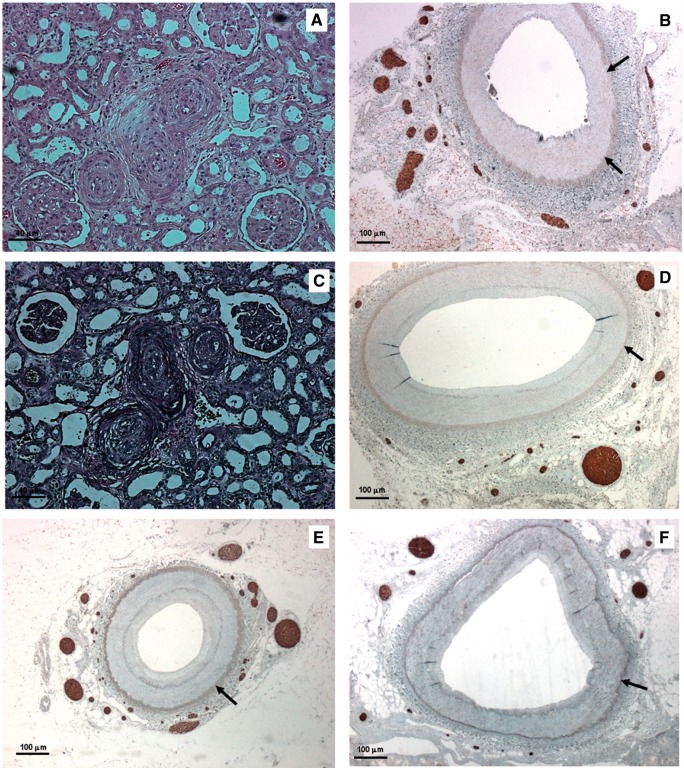
FIGURE 2.
Histologic findings of native and transplanted kidneys in Patient 2 (A–D) and Patient 3 (E, F). (A) Renal arterioles of native kidney with near occlusive concentric intimal thickening (score = 4) (haematoxylin and eosin—bar = 40 μm). (B) Native renal artery with numerous nerves within the adventitia (NFP stain—bar = 100 μm). (C) Renal arterioles of transplanted kidney with severe arteriolar damage and concentric elastic duplication (score = 5) (Weigert stain—bar = 40 μm). (D) Transplanted renal artery: complete regeneration of periadventitial nerves was observed with nerve density equalling values of native artery (GAP43 stain—bar = 100 μm). (E) Native renal artery with a significant density of nerves within the adventitia (NFP stain—bar = 100 μm). (F) Transplanted renal artery: complete regeneration of GAP43-positive nerves with nerve density similar to that measured in native kidneys (GAP43 stain—bar = 100 μm). (Adventitial-medial borders are highlighted by arrows.)

