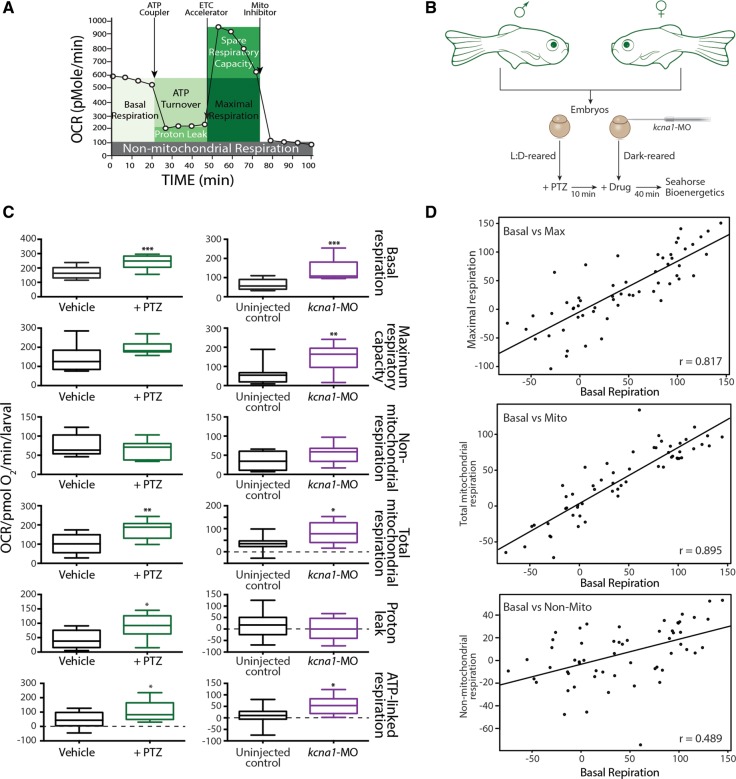
Figure 1.
Metabolic characterization of PTZ and kcna1-MO models of epilepsy. (A) Cartoon representation of how the Seahorse bioanalyser displays mitochondria bioenergetics as modulated by pharmacological inhibitors. (B) Schematic representation of treatment paradigm for metabolic drug screen in PTZ and kcna1-MO models of epilepsy. (C) PTZ and kcna1-MO models of epilepsy exhibit significant increases in metabolic parameters. Specifically, changes in basal respiration, maximal respiratory capacity, non-mitochondria respiration, total mitochondrial respiration, proton leaks, and ATP-linked respiration were studied for both models. Non-mitochondrially mediated changes in respiration were not observed in either model. (D) Linear regression analyses showing significant correlation between basal respiration and both maximum respiratory capacity (r = 0.817; P < 0.0001) and mitochondria-mediated respiration (r = 0.895; P < 0.0001) but only a moderate correlation with non-mitochondrial respiration (r = 0.489; P < 0.001). Data in C are shown as mean ± SEM; *P < 0.05, **P < 0.001, ***P < 0.0001 (unpaired t-test); n = 6–7 fish per group.

