
Figure 2.
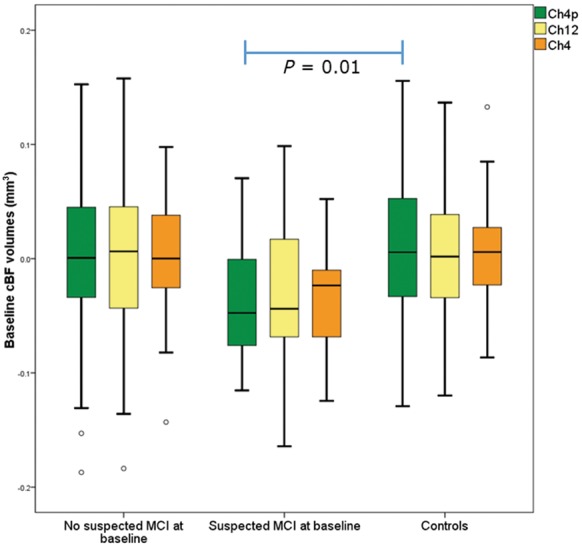
Box plot of cBF region of interest volumes at baseline in controls and Parkinson’s disease patients with and without suspected MCI. CBF region of interest volumes in controls, patients with Parkinson’s disease with and without suspected MCI, with the Ch4p region (corresponding to the posterior NBM) being significantly different between controls and Parkinson’s disease patients with suspected MCI.