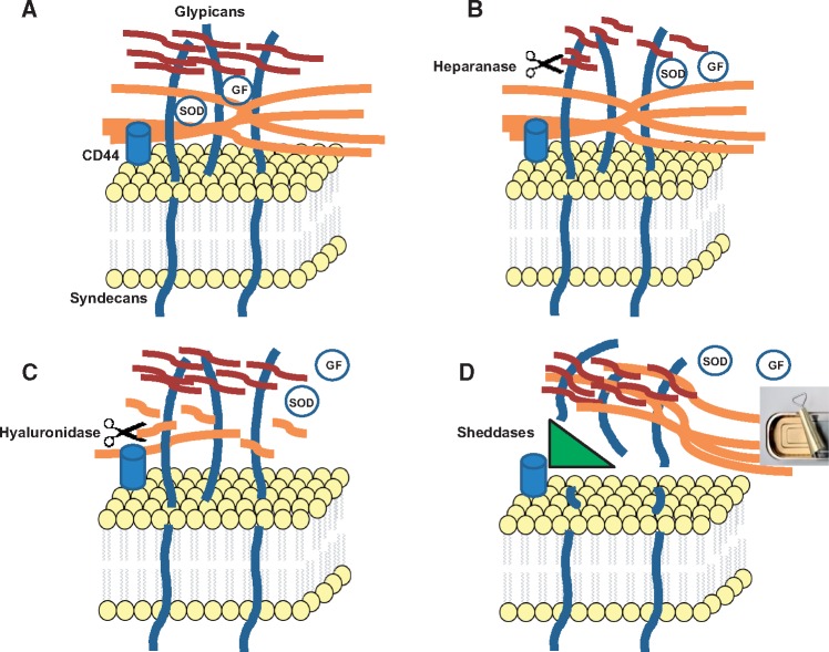
FIGURE 1.
Scenarios for degradation of EG. (A) A simplified schema of EG. HA (orange); heparan sulfate (red; dermatan and chondroitin sulfates are not shown for clarity). Note that diverse proteins are nested in EG, as those shown, as well as albumin and others (see text). GF, growth factors. (B) Heparanase-induced fragmentation of HS results in a partial degradation of EG. (C) Hyaluronidase-induced breakdown of HA chains results in a partial degradation of EG and an increase in permeability. (D) Sheddases cleave proteoglycans, syndecans and glypicans, resulting in peeling off of the entire EG structure. It should be noted that derangements in each component of EG lead in time to disorganization of other components. Hence, the schema depicts only the initial action of EG-degrading enzymes. Nonetheless, this latter scenario based on the action of sheddases results in a most severe and complete degradation of EG (akin to opening a can).

