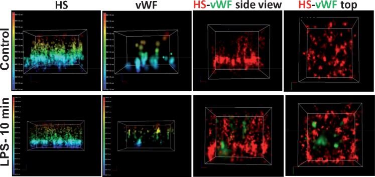
FIGURE 3.
Representative STORM-acquired images of anti-HS-labeled EG and vWF distribution in controls and 10 min after LPS. The panels show three-dimensional reconstructed images of cultured endothelial cells stained with antibodies against HS and vWF. Note that preparations were nonpermeabilized so that vWF readily detectable after LPS is located extracellularly. Images display the height of EG and detectability of vWF in controls and 10 min after application of LPS (left panels) and side or top views. Note the readily detectable appearance of vWF 10 min after LPS, where it encroaches into the EG. The sites of detectable vWF also appear to be depleted of HS.

