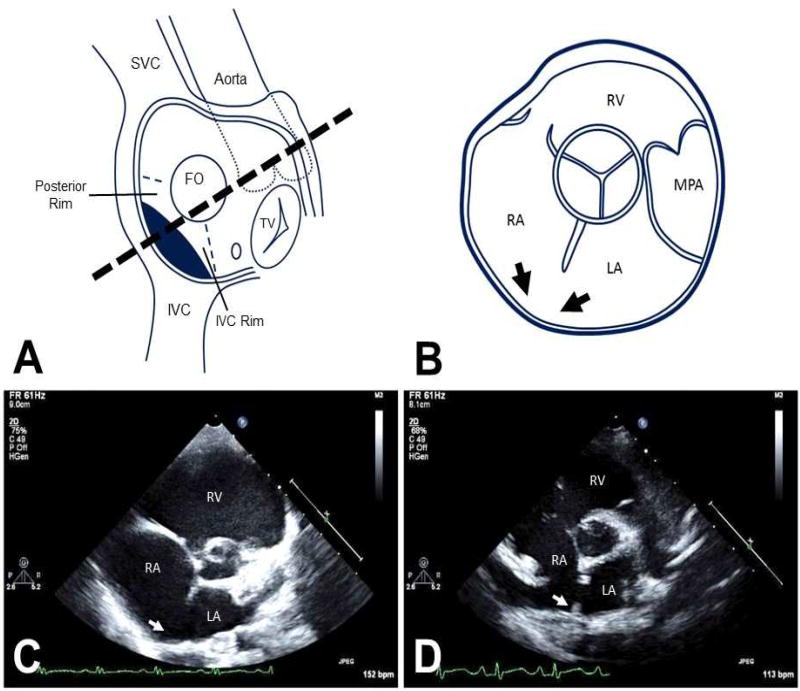
Figure 1.
Anatomy and corresponding parasternal short axis views of the atrial structures. A: The components of the atrial septum are depicted in an en-face view, highlighting the posterior and IVC rims. FO=fossa ovalis part of atrial septum is depicted by a circle. The position of the inferior SVD is shown by the dark shading. B: Cartoon of the corresponding parasternal short-axis view of TTE. The absent posterior rim of the atrial septum creating the "bald" posterior wall of the left atrium, is depicted by the two black arrows. C: Parasternal short axis view demonstrating absence of the posterior rim (white arrow) in a typical inferior SVD. D: Similar parasternal short axis view showing a secundum ASD with the presence of a tiny posterior rim (white arrow). IVC=inferior vena cava, LA=left atrium, MPA=main pulmonary artery, RA=right atrium. RV=right ventricle, SVC=superior vena cava, TV=tricuspid valve.

