FIG. 1.
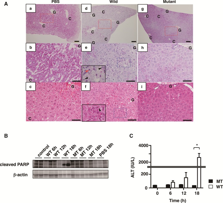
Characterization of livers from mice treated with Cholix. A, Representative liver images stained by Periodic acid-Schiff: PAS (a, b, d, e, g, h) or hematoxylin–eosin: HE (c, f, i) from mice treated with PBS control (a-c), wild-type Cholix (d–f), and mutant-type Cholix (g–i) for 24 h. b, e, and h show high-magnification views of the red rectangular areas in a, d, and g, respectively. Paired images of b/c, e/f and h/i are the same areas stained by PAS/HE with serial sections, respectively. Insets in e or f show high-magnification views of damaged liver with apoptotic cells (arrowheads) and granulocytes (red arrowhead) in the blue rectangular areas of e or f respectively. Scale bars = 100 μm (a, d, c) and 50 μm (b, c, e, f, h, i). C and G, indicate central vein and Glisson’s sheath, respectively. B and C, Time-course of Cholix-injected mice. Mice were injected I.P. with PBS (n=3), wild-type Cholix (n=4), and mutant Cholix(E581A) (n=3). Liver and serum were obtained from each mouse at the indicated times, and then PARP cleavage in the liver (B) and serum ALT level (C) were determined as described in Materials and Methods section. Two representative samples from three mice of each group were utilized for immunoblotting. C, Average and standard deviations of serum ALT levels in each group is shown and significance is *P <0.01. The black bar indicates mutant Cholix(E581A) (MT) and white bar is wild-type Cholix (WT).

