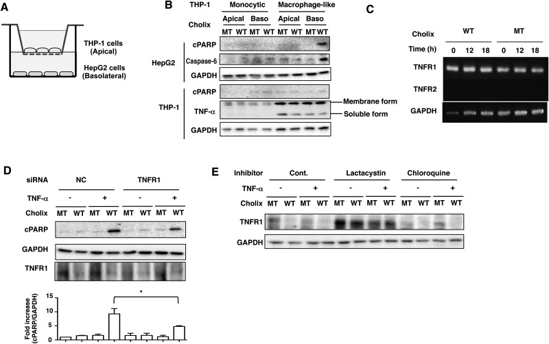
FIG. 4.
Apoptotic signals are induced by Cholix in the co-culture system. A, Schematic drawing of co-culture system. Confluent THP-1 cells or PMA-differentiated macrophage-like cells were plated on apical side (Apical), which has a semipermeable membrane. HepG2 cells (1 × 10⁴ cells/well) were plated on basolateral side (Baso) and the system was cultured overnight. B, Cholix (10 μg/ml) was added to the apical or basolateral media and the cells were incubated for 12 h and then collected. Then, cells were lysed with 1×SDS sample buffer for immunoblotting with the indicated antibodies. Experiments were repeated three times with similar results. C, HepG2 cells were incubated with mutant or wild-type Cholix for 0, 12, 18 h, and then total RNA was extracted. Expression of TNFR1, TNFR2, and GAPDH mRNA was detected by RT-PCR as described in Materials and Methods section. D, The indicated siRNA-transfected HepG2 cells were incubated for 12 h with 10 μg/ml wild-type Cholix (WT) or mutant Cholix(E581A) (MT)in the presence or absence of TNF-α (25 ng/ml). Then, cells were lysed with 1×SDS sample buffer for immunoblotting with the indicated antibodies. Experiments were repeated three times with similar results. Quantification of TNF-α/Cholix-induced PARP cleavage was performed by densitometry (bottom panel). Data are presented as mean ±SD of values from three experiments and significance is *P<0.05. E, Cells were treated for 30 min with or without 10 μM lactacystin or 100 μM chloroquine, and then incubated for 12 h with 10 μg/ml wild-type Cholix (WT) or mutant Cholix (MT) in the presence or absence of TNF-α (25 ng/ml). Cells were lysed with 1×SDS sample buffer for immunoblotting with the indicated antibodies. A blot representative of three separate experiments is shown.

