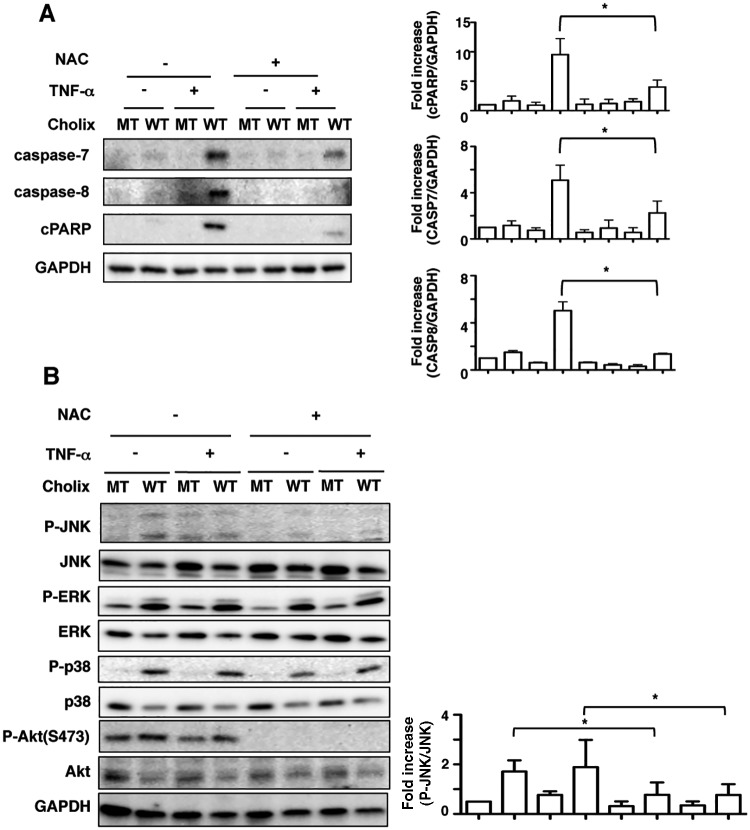
FIG. 7.
ROS inhibitor suppresses TNF-α/Cholix-induced apoptotic signaling. A, Cells were pretreated for 30 min with or without 10 mM NAC, and then incubated for 12 h with wild-type Cholix (WT) or mutant Cholix(E581A) (MT) in the presence or absence of TNF-α. Then, cells were lysed with 1×SDS sample buffer for immunoblotting with the indicated antibodies. A blot representative of three separate experiments is shown. Quantification of TNF-α/Cholix-induced caspase-7, -8, and PARP cleavage levels in HepG2 cells was performed by densitometry (right panel). Data are presented as mean ±SD from three experiments and significance is *P<0.05. B, Cells were pretreated for 30 min with or without 10 mM NAC, and then incubated for 12 h with wild-type Cholix (WT) or mutant Cholix(E581A) (MT) in the presence or absence of TNF-α. Then, cells were lysed with 1×SDS sample buffer for immunoblotting with the indicated antibodies. A blot representative of three separate experiments is shown. Quantification of phospho-JNK level in HepG2 cells was performed by densitometry (right panel). Data are presented as mean ±SD from three experiments and significance is *P<0.05.

