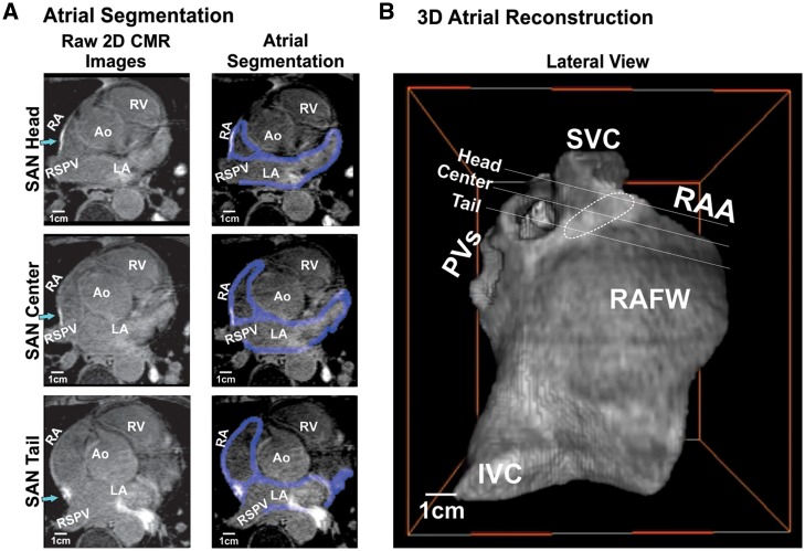
Figure 1.
In vivo LGE-CMR segmentation and 3D atrial reconstruction. (A) 2D imaging cross sections from 3D LGE-CMR SAN head, centre, and tail sections of Volunteer 1A with surrounding atrial tissue. Left: 2D raw images, green arrows point to the SAN head, centre, and tail, respectively; Right: atrial wall segmentation shown in blue. (B) 3D atrial reconstruction of the 2D imaging cross sections of Volunteer 1A. Ao, aorta; IVC, inferior vena cava; PVs, pulmonary veins; RA, right atria; RAA, right atrial appendage; RSPV, right superior pulmonary vein; RV, right ventricle; SAN, sinoatrial node; SVC, superior vena cava.

