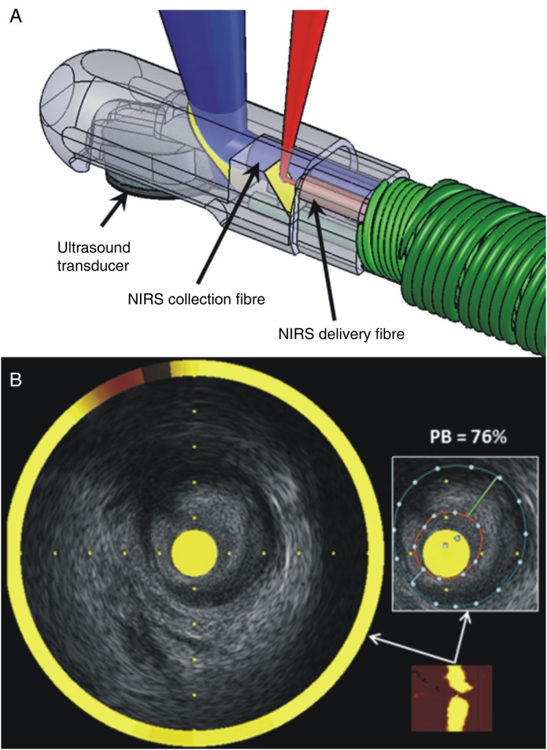
Figure 2.

Combined near infrared spectroscopy—intravascular ultrasound catheter. (A) The tip of the catheter incorporates a rotating intravascular ultrasound transducer operating at 50 MHz with extended bandwidth, and two near infrared spectroscopy fibres that transmit and collect the near infrared light. (B) The chemogram is the output of the near infrared spectroscopy catheter (bottom right), is co-registered with the intravascular ultrasound data creating hybrid images that allow assessment of lumen, outer vessel wall, and plaque dimensions including plaque burden and simultaneous evaluation of the longitudinal and circumferential distribution of the lipid component. Panel B was reprinted with permission from Madder et al.35
