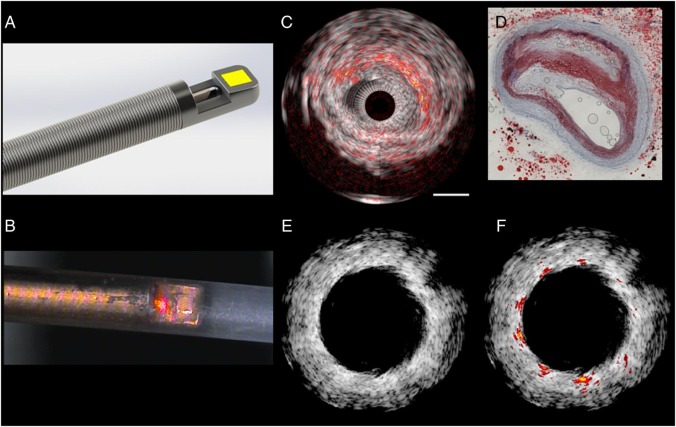
Figure 6.
Intravascular ultrasound - intravascular photoacoustic imaging. (A) Sketch of a catheter tip, showing the ultrasound transducer (yellow) aligned with the tip of a side-looking optical fibre on a flexible drive shaft. (B) Microphotograph of an experimental catheter device (figure provided by M. Wu and G. Springeling unpublished), with a red pointer laser indicating the optical channel, in a polymer sheath (outer diameter 1.1 mm). (C) Lipid imaging of a human atherosclerotic plaque ex vivo, wavelength = 1710 nm. Conventional intravascular ultrasound is shown in greyscale, with intravascular photoacoustic lipid signal in red-orange overlay. Comparison with histology (D; Oil Red O stain) shows high intravascular photoacoustic signal in lipid-rich areas. (E) Stent imaging with intravascular ultrasound in an atherosclerotic vessel with highly echogenic plaque. The stent struts provide very limited contrast. (F) The high intravascular photoacoustic signal generated by the metal stent struts allows accurate assessment of stent apposition (J. Su, unpublished).

