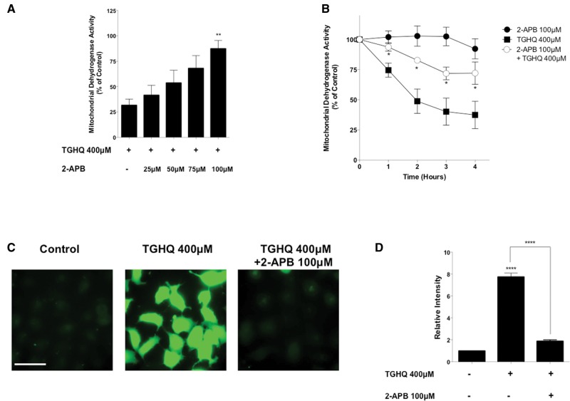
Figure 2.
SOCs facilitate increases in intracellular calcium. (A) HK-2 cells were pre-treated for 30 min and co-treated with SOC inhibitor 2-APB at various concentrations (25–100 μM) in the presence or absence of TGHQ 400 μM for 2 h. (B) HK-2 cells were pre-treated for 30 min and co-treated with 2-APB (100 μM) in the presence of TGHQ 400 μM for various periods of time (1–4 h). Cell viability was determined using the MTS based assay. (C) Cells were incubated with Fluo-4 AM (5 µM) and pre-treated with 2-APB (100 μM), then co-treated with TGHQ 400 μM for 2 h. Cells were immediately imaged using ×40 magnification under a deconvolution microscope. Scale bar 100 µm. (D) The densitometric and statistical analysis of the calcium imaging data. All data are mean ± SE; n ≥ 3. *P < .05, **P < .01 when compared with TGHQ by one-way ANOVA followed by a post-hoc Tukey’s test. ****P < .001 when compared with control by one-way ANOVA followed by a post-hoc Tukey’s test.

