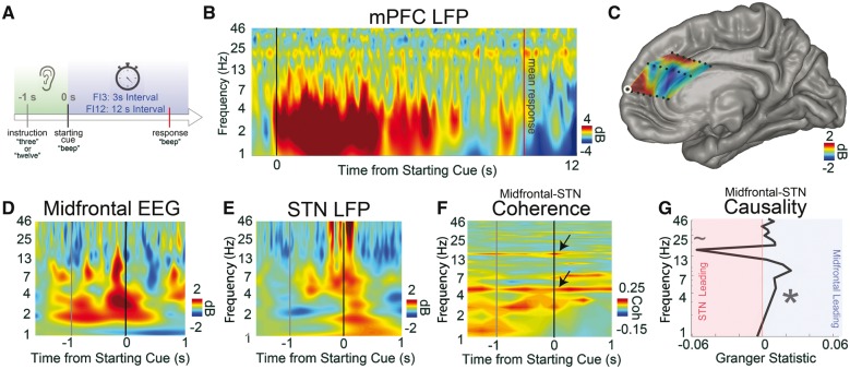
Figure 3.
During cognitive processing, 4-Hz medial PFC signals exert top-down control of the STN. (A) We used interval timing tasks to study elementary cognitive processing in PFC and STN. During interval timing, patients heard an instruction telling them how long to wait (3 or 12 s), then a starting cue indicating that they should start timing. Patients reported their estimate of the interval by making a button-press response. (B) We recorded LFPs from depth electrodes in human medial PFC (mPFC), which has been implicated in interval timing across species. Time-frequency analysis revealed robust cue-triggered low-frequency delta/theta activity between 1 and 8 Hz in medial PFC. (C) The amount of 1–4 Hz activity at 300–500 ms post-cue was interpolated across medial PFC depth electrodes, showing cue-triggered modulation at dorsal medial PFC and rostral and middle cingulate gyri. (Contact from B outlined in white.) (D and E) We collected human intraoperative data from 10 patients with Parkinson’s disease undergoing STN-DBS implantation performing the interval timing task. Time-frequency analyses revealed cue-triggered delta/theta activity from intraoperative midfrontal EEG (∼AFz/Fz) and STN during cues. (F) Midfrontal EEG and STN had strong, task-independent coherence at ∼4 Hz and at ∼20 Hz in the beta range (arrows; raw coherence >0.17 is significant at 95% CI). (G) Granger causality analyses around the cue revealed that at 4 Hz, midfrontal EEG oscillations led the STN while ∼20-Hz STN oscillations led midfrontal EEG. Ten patients with Parkinson’s disease undergoing STN-DBS implantation. *P < 0.05; ∼P < 0.10. All data from FI12 trials. See also Supplementary Fig. 4.

