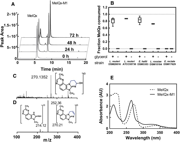
Figure 1.
Detection and characterization of MelQx metabolite (MelQx-M1) in bacterial culture. A, Presence of MelQx and MelQx-M1 during growth of E. hallii DSM 3353 in mYCFA containing 0.8% (v/v) glycerol (approximately 100 mM) and 100 µM MelQx, at 37 °C. B, Degradation of MelQx by L. reuteri DSM 20016 and ATCC 55730, E. hallii DSM 3353, L. rossaie DSM 15184 and E. rectale DSM 17629 in the presence and absence of glycerol in mYCFA medium. Data are displayed as mean values ± SD from 3 independent experiments. C, high resolution mass spectra of peak MelQx-M1 showed a m/z 270.1352. D, fragmentation of m/z 270.1352 at 8.5 min of the sample 72-h MelQx-incubation. E, UV absorbance of MelQx (dashed line) and MelQx-M1 (solid line).

