Figure 1.
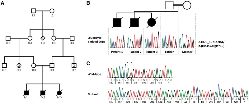
DNA and RNA analysis in the family with three siblings carrying the homozygous ATAD1 mutation. (A) Pedigree of the family. (B) Partial sequence electropherograms demonstrating the ATAD1 c.1070_1071delAT [p.(His357Argfs*15)] mutation in the homozygous state in leucocyte-derived DNA of the affected siblings (Patients 1–3). Their healthy parents (father and mother) are heterozygous carriers of the mutation. (C) Partial sequence electropherograms show the 2-bp deletion in ATAD1 in fibroblast-derived cDNA of one sibling (Mutant) in comparison to the cDNA sequence of a healthy individual (Wild-type). Deleted bases are marked by parenthesis in the normal sequence. The encoded amino acid residues are depicted below each sequence in the three-letter code and show the 14 novel amino acid residues at the C-terminus of ATAD1 (highlighted in bold). Asterisk indicates a stop codon.

