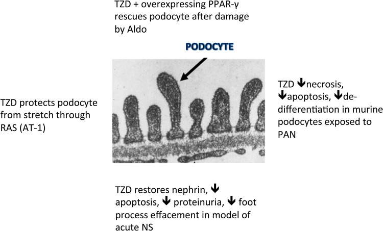
FIGURE 6.
TZD podocyte-protective effects explained. After stretch: through RAS blockade [72]. In PAN nephritis: through restoration in balance of pro-apoptotic (caspase 3) and anti-apoptotic (Bcl-xl) molecules and reduction in pro-inflammatory TGF-β expression [11]. After damage by aldosterone [70]: overexpression of PPAR-γ/use of rosiglitazone, rescues podocytes through decreased ROS and maintenance of cell morphology (restores nephrin expression). Both are on-target effects (blocked by small interfering PPAR-γ RNA). In acute nephrosis [65], podocytes cause a decrease in the expression of nephrin, phosphorylated Akt and α-actinin 4 and an increase in apoptosis. PPAR-γ agonists given around the time of the injury produce a reversal of these effects as well as a reduction in proteinuria, a decrease in desmin and an improvement in foot process effacement [63].

