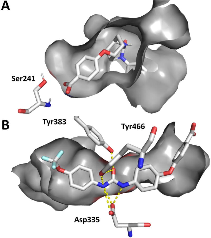
Figure 3.
A. Docking of t-TUCB (yellow) in the active site of FAAH (PDB: 1MT5) using AutoDock Vina and B. Co-crystal structure of t-TUCB (yellow) in the active site of sEH. The key catalytic residues for FAAH (Ser241) and sEH (Asp335) are represented in addition to the proton-donating residue on sEH (Tyr 383 and Tyr466). Hydrogen bonds are represented on the co-crystal structure as yellow dashed lines.

