
Fig. 1. Size and morphology of milk exosomes.
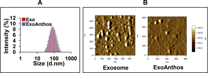
Bovine milk-derived exosomes (Exo) and ExoAnthos formulation were analyzed for size by zetasizer (A). Diluted Exo and ExoAnthos suspensions were used for imaging by AFM. Topographic and amplitude images were captured concurrently with a fixed force (<1 nN) with a scanning rate of 1 Hz (B).