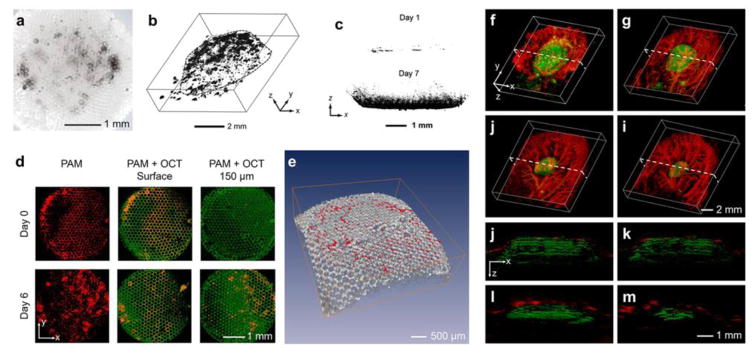
Figure 2. Interrogating biomaterial-tissue interactions.
(a) Bright-field optical image showing melanoma cells grown in a porous PLGA scaffold. Adapted with permission from [38]. (b) PAT reconstruction image showing 3D distribution of melanoma cells in a PLGA scaffold. Adapted with permission from [66]. (c) PAT images showing invasion of NIH/3T3 fibroblasts into PLGA scaffolds at days 1 and 7; the cells were stained with formazan to generate exogenous contrast. Adapted with permission from [43]. (d) Top view of the PAT/OCT images showing the ingrowth of melanoma cells from the surface into the center of the PLGA scaffolds. The melanoma cells were imaged by the PAT subsystem whereas the scaffold by the OCT subsystem, both in a label-free manner. (e) Volumetric rendering of the OCT-imaged scaffold (gray) with PAT-imaged melanoma cells (red). Adapted with permission from [38]. (f–i) Co-registered 3D reconstruction PAT images showing both the degradation of an individual PLGA scaffold and the remodeling of vasculature simultaneously in vivo. (j–m) Co-registered cross-sectional PAT images at the dotted planes as indicated in (f–i), respectively. Adapted with permission from [44].

