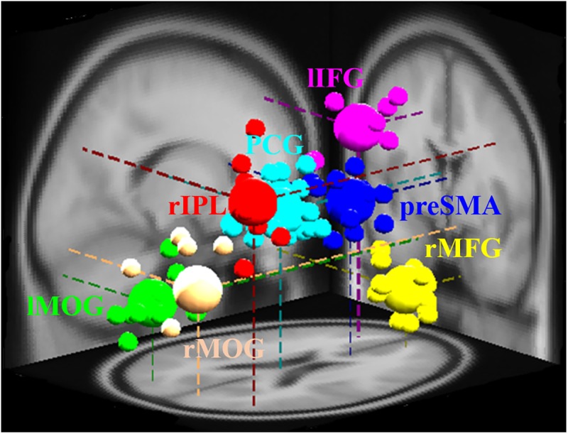
FIGURE 2.

Different brain regions and related dipole source locations. Pre-SMA, pre-supplementary motor area; rMFG, right middle frontal gyrus; lIFG, left inferior frontal gyrus; PCG, posterior cingulate gyrus; rIPL, right inferior parietal lobe; lMOG, left middle occipital gyrus; rMOG, right middle occipital gyrus. Pre-SMA, lIFG, and rMFG are accomplished for response inhibition. PCG and rIPL are accomplished for emotion and motivation. Bilateral MOGs are accomplished for the analysis of visual stimuli. The small area shows the location of each participant’s dipole, while vast areas display the dipole locations of each cluster.
