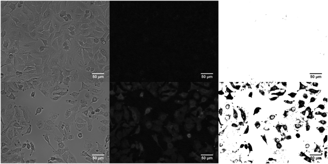
Figure 5.
Fluorescent microscopy was performed for cell transfection visualisation. Cells with pMaxGFP and no electrospray (a–c). Electrospray mediated pMaxGFP transfection (d–f). Image processing was performed using Fiji image processing. Electrospray was performed with sucrose-based media with osmolarity of 370 mOsm. Working distance of 4 mm, flow rate of 20 µl/min with 3 kV voltage. 10 min incubation time was used. Analysis was performed after 24 hours.

