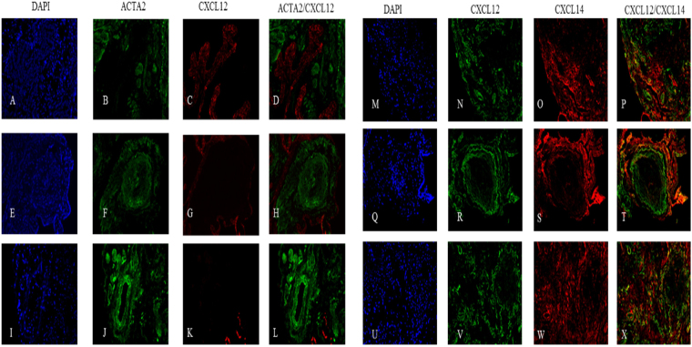
Figure 8.
Histological localization of CXCL12 and CXCL14 in IPF. Representative images of three IPF tissue sections are stained for the presence of CXCL12 (C,G,K,N,R,V), ACTA2 (B,F,J), and CXCL14 (O,S,W). CXCL12 is absent in foci (H,T) or areas of significant fibrosis (L), sparingly distributed in lower fibrosis regions (S), and highly present in airways (D). No co-localization of ACTA2 and CXCL12 is observed (D,H,L), with minimal co-localization of CXCL12 and CXCL14 seen in diffuse fibrosis (P,T,N).

