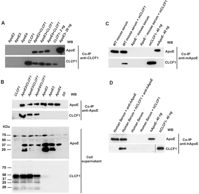
Figure 1.
CLCF1 binds recombinant and serum ApoE. (A) Recombinant CLCF1 (200 ng) and ApoE2, 3 or 4 (250 ng) were incubated alone or in combination for 16 h. The samples were subjected to immunoprecipitation (IP) with anti-CLCF1 and protein G agarose. Immunoprecipitated proteins were analyzed by WB using mAbs specific for ApoE (upper panel) or CLCF1 (lower panel). Lanes “CLC 2 ng” and “ApoE3 20 ng”, show results of recombinant proteins directly subjected to SDS-PAGE and WB blot analysis. (B) CLCF1 and ApoE form a complex when co-expressed in HEK-293 cells. The cell culture medium from stable transfectants expressing the indicated proteins in combination with CRLF1 or from cells transfected with empty expression vector (lane D5) was subjected to immunoaffinity chromatography with rat-anti-human ApoE and anti-rat Ig Agarose. The eluates were analysed by WB using anti-ApoE or anti-CLC mAbs (upper panels). Supernatants of the stable transfectants were analyzed for the presence of ApoE or CLCF1 by WB (lower panels). (C and D) CLCF1 binds mouse and human serum ApoE. (C) Mouse serum samples isolated from WT or ApoE−/− mice were diluted 1:20, mixed with biotinylated mouse CLCF1 and the complexes immunoprecipitated with rabbit anti-mouse ApoE and anti-rabbit Ig agarose. The eluates were analysed for ApoE or CLCF1 by WB using anti-mouse ApoE or HRP-labelled streptavidin. The last two lanes show the signals obtained with 40 ng of biotinylated mouse CLCF1 or ApoE included as control. (D) Human serum samples were diluted 1:20, mixed with biotinylated human CLCF1 and the complexes immunoprecipitated with rat anti-ApoE and anti-rat Ig agarose. The eluates were analyzed for ApoE or CLCF1 by WB using anti-ApoE or HRP-labelled streptavidin. The last two lanes show the signals obtained with 40 ng of biotinylated human CLCF1 or ApoE included as control. Blots’ images where cropped to show relevant areas.

