Fig. 1.
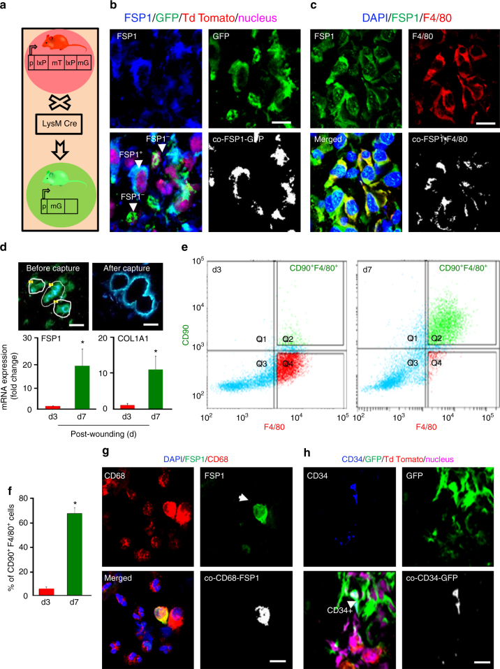
Majority of FSP1+ fibroblast-like cells in wound granulation tissue are of myeloid origin. a LysMCreRosamT/mG mice express cell membrane-localized td Tomato (red) fluorescence while cells of myeloid origin express GFP (green) fluorescence. b FSP1 (blue) immunostaining of LysMCreRosamT/mG mice d5 wound. Colocalization was performed using Olympus Fluoview® software. Estimated (65 ± 5%) FSP1+ cells (blue) were GFP+ demonstrating their myeloid origin, n = 5, scale bar = 10 µm. c Macrophage (F4/80, red) and fibroblast (FSP1, green) immunostaining at wound-edge tissue (d5) from C57BL/6 mice, n = 5, scale bar = 10 µm. d Laser capture micro-dissected (LCM) GFP+ cells from full thickness excisional wounds on d3 (early inflammation phase) or d7 (inflammation resolution phase) post-wounding (PW), scale bar = 10 µm. qRT-PCR of LCM-captured GFP+ cells showed increased expression of FSP1 and Col1A1 at d7, n = 5, Student’s t test p < 0.05. e, f Multicolor flow cytometry of CD11b+ of wound macrophages on d3 and d7 PW with the F4/80 (macrophage, x-axis) and CD90 (fibroblast marker, y-axis) displayed increase (68 ± 2%) in F4/80+CD90+ cells on d7 PW, n = 4, Student’s t test p < 0.05. g Chronic wound fluid derived from human subjects contain CD68+ FSP1+ cells, scale bar = 10 µm. h Fibrocyte marker CD34 (blue), immune-staining of LysMCreRosamT/mG mice d5 wound tissue. Estimated (15 ± 2%) of CD34+ cells (blue) were GFP+, n = 5, scale bar = 10 µm. Data presented as ±SD

