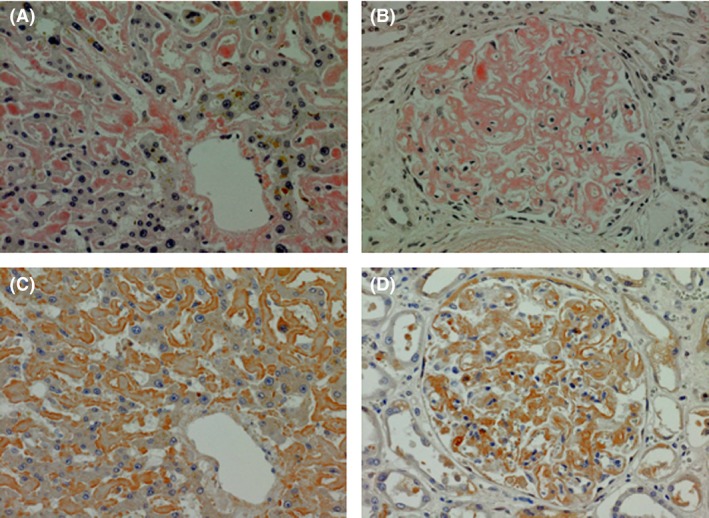
Figure 2.

Autopsy specimens stained with Congo red (A and B) and anti‐Factor X antibody (C and D). (A) Liver, Congo red, amyloid deposition to sinusoid. (B) Kidney, Congo red, amyloid deposition mainly to mesangial matrix. (C and D) Liver and kidney stained with anti‐Factor X, showing that Factor X exists in union with amyloid.
