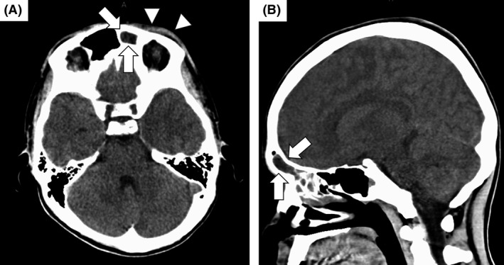
Figure 1.

Computed tomography (CT) images of the head on the day of admission. (A and B) Attenuation of the tissue within the left sinus is observed (arrows). Periorbital soft‐tissue thickening is also observed (arrow heads). Bone abnormalities and continuity with the intracranial space are not observed. Intracranial subdural abscess is not apparent on CT images.
