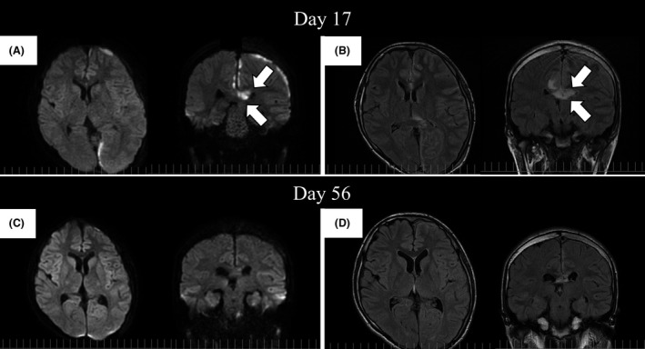
Figure 5.

Follow‐up magnetic resonance imaging studies performed on days 17 and 56. (A) The subdural abscess was remarkably diminished in diffusion‐weighted image (DWI) sequences. However, a high‐intensity area appeared in the corpus callosum (arrows) on day 17. (B) In fluid‐attenuated inversion recovery (FLAIR) sequences, the corpus callosum also exhibited high intensity (arrows), and its involvement in the infection was suspected. (C and D) The corpus callosum lesion remarkably improved in both DWI and FLAIR sequences on day 56.
