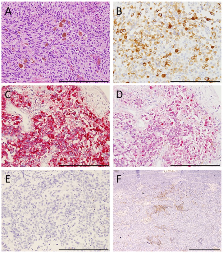
Figure 2.
Histopathological findings. (A) Tumor cells stained with hematoxylin and eosin. (B) Immunohistochemical staining of tumor cells was positive for melan-A, (C) HMB-45, and (D) S-100 and negative for cytokeratin markers, (E) AE1/AE3 [(A-E) original magnification, ×400. Scale bar, 200 µm]. (F) Immunohistochemical membranous positive staining of PD-L1 in tumor cells (original magnification, ×100; scale bar, 500 µm.

