Figure 1.

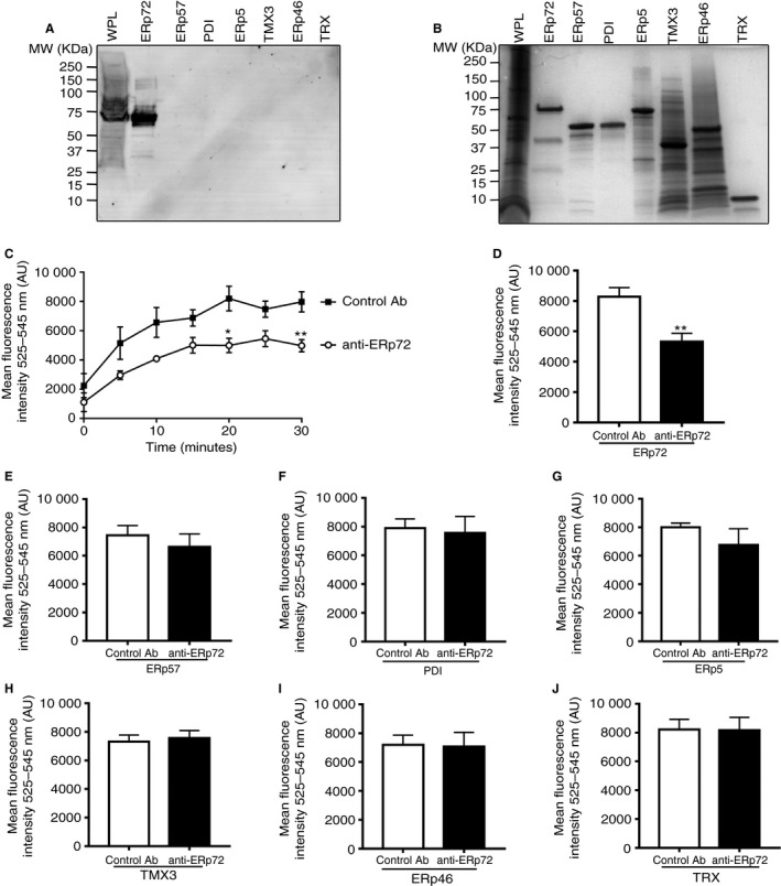
Anti‐ERp72 selectively inhibits ERp72 enzyme activity. Human whole platelet lysate (8 × 10⁸ cells mL −1) and recombinant ERp72, ERp57, PDI, ERp5, TMX3, ERp46 and TRX (all 5 μg) were separated by reducing SDS PAGE and immunoblotted with (A) anti‐ERp72 (1 μg mL −1 diluted in 2% w/v BSA/TBS‐T) and FITC‐conjugated anti‐human secondary antibody (1 : 1000 dilution in 2% w/v BSA/TBS‐T) or (B) stained with Coomassie blue protein stain to reveal total protein loading. (C) Thiol isomerase activity of ERp72 was measured by fluorescence‐based thiol isomerase assay. Anti‐ERp72 (open circles) or control antibody (closed squares, both 25 μg mL −1) were incubated with recombinant ERp72 (100 nm) for 5 min, prior to the addition of DTT (0.5 μm) and DI‐E‐GSSG substrate. Fluorescence values were recorded at 5‐min intervals for a total of 30 min. (D) Inhibition of ERp72 enzyme activity, (E) ERp57 activity, (F) PDI activity, (G) ERp5 activity, (H) TMX3 activity, (I) ERp46 activity or (I) TRX activity at 30 min in the presence of control antibody (open histogram) or anti‐ERp72 (closed histogram) (n = 5). Graphs represent mean ± SEM. P values were calculated by one‐way anova (C) or Student's t‐test (D–J), (*P < 0.01, **P < 0.01).
