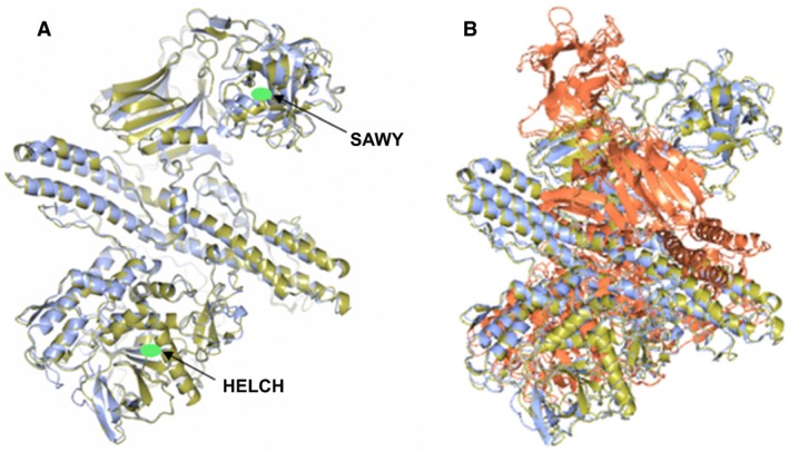
Figure 3.

Structure prediction and comparison of putative eBoNT/J toxin and its associated NTNH to BoNT/A and BoNT/A complexed with its NTNH, respectively.(A) Superposition of the predicted structure of putative eBoNT/J (gold) with the crystal structure of an inactive BoNT/A (blue Protein Data bank ID: 3V0C). Positions of the zinc‐binding site (HELCH) and target cell‐binding motif (SAWY) are indicated by green ovals. (B) Superposition of the predicted structure of the eBoNT/J NTNH (gold) with that determined for BoNT/A (red) complexed with its own NTNH (blue; Protein Data bank ID: 3V0B).
