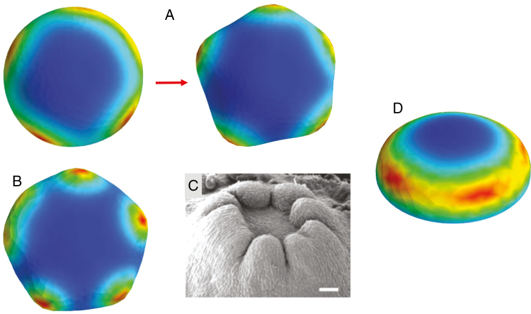
Fig. 5.
Variation in patterns. The model can generate some of the anomalous morphologies seen experimentally. (A) Concentration peak splitting, in this case from an earlier pattern of four peaks (left) to a later pattern of five peaks (right), corresponds to readjustments seen experimentally, where additional cotyledons are sometimes seen a week after the earliest count. (B) Peak fusions, where a ring with space for, in this case, between five and six peaks, has several peaks ‘smeared’ together, corresponding to fused or extra-width cotyledons sometimes observed experimentally (C; from Harrison and von Aderkas, 2004, with permission; scale bar = 250 µm). Such cases of indistinct peak number tend to occur at transitional radii between distinct integer peak numbers (i.e. between the shapes shown in Fig. 4). (D) Transient pattern in early stages of an ‘NPA-treatment’ simulation (at later stages, this passive P2 pattern is distributed in a smooth ring). Such transient pattern could correspond to the ‘bumpy cup’ morphology sometimes observed in NPA-treated embryos, where the cup rim is not smooth (Holloway et al., 2016).

