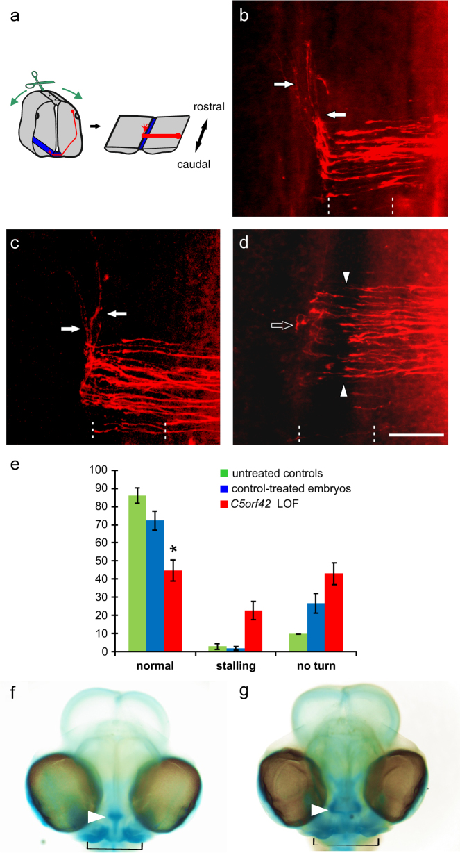
Fig. 4.
Silencing C5orf42 in the developing neural tube of chicken embryos resulted in pathfinding errors of commissural axons at the midline. The pathfinding behavior of dI1 commissural axons at the floor plate was analyzed in open-book preparations of the spinal cord (a; see Methods section for details). Axonal trajectories (red) are visualized by the injection of DiI into the area of commissural neuron cell bodies. In untreated control embryos, commissural axons grew ventrally, entered the floor plate to cross the midline and turned rostrally at the floor-plate exit site (arrows, b). No difference in commissural axon pathfinding was observed in control-treated embryos (c). In contrast, after silencing C5orf42 in the neural tube, dI1 commissural axons failed to cross the midline, stalled in the floor plate (arrow heads) and also failed to turn rostrally along the contralateral floor-plate border (open arrow, d). The floor plate is indicated by dashed lines. Bar: 50 µm. Quantification of phenotypes as average percentage of DiI injection sites per embryo with the given phenotypes (e, shown with SEM; *P < 0.05). Green bars for untreated control embryos, blue bars for control-treated embryos-expressing GFP, red bars for embryos after RNAi-mediated silencing of C5orf42 (LOF, loss-of-function). In addition, silencing C5orf42 in cranial neural crest cells of developing chicken embryos resulted in facial dysmorphism. In comparison with the control-treated embryos (f), embryos lacking C5orf42 in cranial neural crest cells developed aberrant facial features (g). The nasal structure (arrowhead) was much wider than in age-matched control heads stained with Alcian Blue. Similarly, the jaw was broader in experimental compared to control embryos (indicated by black line) and eye distance was increased

