Fig. 1.
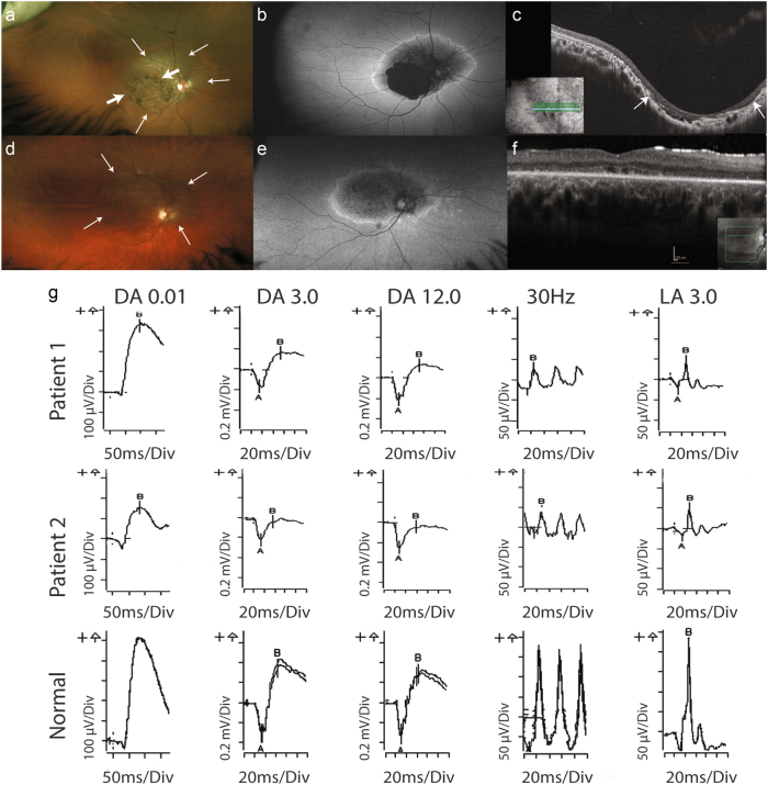
Ophthalmic investigations demonstrating macular atrophy with cone dystrophy in Case 1 and cone-rod dystrophy in Case 2. Patient 1—right eye (a–c): a Wide-field colour-corrected fundus images illustrating the coloboma-like macular atrophy with thick arrows marking the boundaries, and surrounding retinal atrophy extending beyond the vascular arcades and nasal to the optic discs with thin arrows marking this change. b Wide-field fundus autofluorescence showing the complete loss of autofluorescence coinciding with the coloboma-like macular atrophy surrounded by blotchy hypoautofluorescence and the ring of hyperautofluorescence bounding the atrophic area. c Optical coherence tomography (OCT) images showing the excavation and loss of the photoreceptor layers including the ellipsoid layer at the site of the macular atrophy, which is demarcated by the arrows. Patient 2—right eye (d–f): d Wide-field colour- corrected fundus images illustrating the macular atrophy with surrounding retinal atrophy extending beyond the vascular arcades and nasal to the optic discs, with thin arrows marking the extent of the atrophy. e Wide-field fundus autofluorescence showing blotchy hypoautofluorescence in the posterior pole and the ring of hyperautofluorescence bounding the atrophic area. f OCT images showing the loss of the photoreceptor ellipsoid layer at the site of the macular atrophy indicated by the arrows. g Full-field electroretinogram (ERG) performed according to ISCEV standards. Trace results for the right eye in patients 1 and 2 compared with normal. Patient 1 shows cone photoreceptor dysfunction with reduction in the photopic ERG indicated by reduction in the light-adapted (LA) 30 Hz flicker and LA 3.0 stimulus tests. There is a mild reduction in the rod-mediated scotopic ERG under the following stimulus conditions: DA0.01, DA3.0 and DA12.0. Patient 2 shows cone photoreceptor dysfunction with a reduction in the photopic ERG indicated by reduction in the 30 Hz flicker and LA 3.0 stimulus tests. There is a moderate reduction in the rod-mediated scotopic ERG under the following stimulus conditions: DA0.01, DA3.0 and DA12.0

