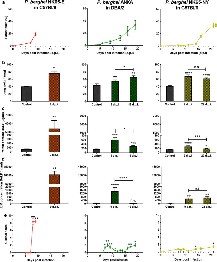
Fig. 1.

P. berghei NK65-E-infected C57BL/6 mice developed more severe lung pathology compared with P. berghei ANKA-infected DBA/2 mice and P. berghei NK65-NY-infected C57BL/6 mice. C57BL/6 mice were infected with P. berghei NK65-E or P. berghei NK65-NY and DBA/2 mice were infected with P. berghei ANKA. a Peripheral parasitaemia levels were determined through blood smears at the indicated time points (n = 7–32 per group). Mice were sacrificed at the indicated times and lung weight b, protein content c and IgM concentration d in BALF were measured (control: n = 4–10 per group, infected: n = 7–23 per group). e Clinical score was monitored throughout the course of infection (control: n = 4–8 per group, infected: 8–20 per group). Asterisks on top of the bars indicate significant differences compared to the uninfected control group. Horizontal lines with asterisks on top indicate significant differences between groups and horizontal lines with n.s. on top indicate no significant differences between groups
