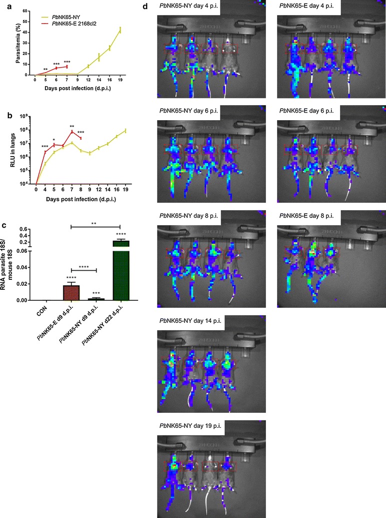
Fig. 5.

Tissue tropism of the P. berghei NK65-NY and P. berghei NK65-E parasite. C57BL/6 mice were infected with P. berghei NK65-NY (green, PbNK65-NY) or P. berghei NK65-E 2168cl2 (red, PbNK65-E). a Peripheral parasitemia was determined through flow cytometry at indicated time points. b The accumulation of P. berghei NK65 schizonts in lungs was measured and visualized through the bioluminescent features of the GFP-luc transfected parasites at the indicated time points and expressed as relative light units (RLU) (n = 8 per group). Asterisks indicate significant differences between groups. c C57BL/6 mice were infected with P. berghei NK65-NY or P. berghei NK65-E, and parasite loads in perfused large lungs were determined by quantifying parasite 18S RNA transcripts by RT-qPCR (n = 8–11 per group). Asterisks on top of the bars indicate significant differences compared to the uninfected control group. Horizontal lines with asterisks on top indicate significant differences between groups. d Whole-body bioluminescence images of representative infected mice are shown
