
FIG 1.
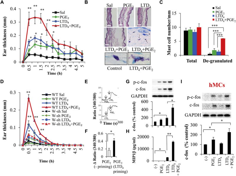
Effect of LTD4 and PGE2 on ear edema in mice in vivo and MC activation in vitro. Wild-type (WT) BALB/c mice were treated with saline (Sal), 0.5 μmol/L LTD4, 0.5 μmol/L PGE2, or LTD4 plus PGE2. A, Ear thickness. B, Hematoxylin and eosin staining and toluidine blue staining. C, Quantification (blind analysis) of MCs per millimeter. D, Ear thickness in C57BL/6 and W-sh mice treated with 0.5 μmol/L LTD4, PGE2, and LTD4 plus PGE2. Results are means ± SEMs from 4 to 6 mice per group per experiment and 3 experiments performed. E–I, LAD2 cells (Fig 1, E–H) and hMCs (Fig 1, I) were stimulated with 0.5 μmol/L LTD4, 0.5 μmol/L PGE2, or both, and calcium flux (Fig 1, E and F), c-fos (Fig 1, G and I), and MIP-1β (Fig 1, H) were analyzed. *P < .05, **P < .01, and ***P < .001.