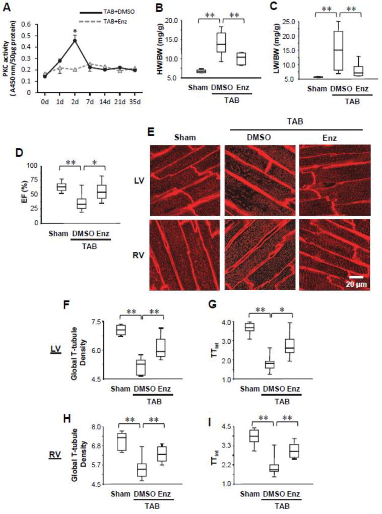
Figure 1. PKC inhibition protects against cardiac dysfunction and T-tubule remodeling following pressure overload.
A, Transient activation of PKC in mouse hearts induced by pressure overload. Administration of PKC inhibitor enzastaurin (Enz) abolished the transient activation of PKC. B–D, Summary data of heart weight/body weight ratio (HW/BW, B), lung weight/body weight ratio (LW/BW, C), and ejection fraction (EF, D) 5 wks after TAB or sham surgery. Some TAB mice were administered Enz, which attenuated the development of heart failure. n ≥ 6 mice/group. E, Representative left ventricular (LV) and right ventricular (RV) in situ confocal T-tubule images after staining with lipophilic marker MM4–64. F–I, Summary data in LV (F, G) and RV (H, I) of global T-tubule density (F, H) and the index of T-tubule integrity (TTint) (G, I). n=6, 10, 6 hearts for sham, TAB+DMSO and TAB+Enz, respectively. For each heart, 10 images were recorded from LV and RV, respectively. The analyzed outputs from the same heart were averaged to represent one heart. * p<0.05, ** p <0.01.

