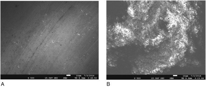
Figure 3.

Scanning electron micrographs of titamium (A) and hydroxyapatite (B) discs used for dental biofilm formation. Notable differences can be observed with regards to their relative microtopography HA showing a much more roughened surface (×500).
