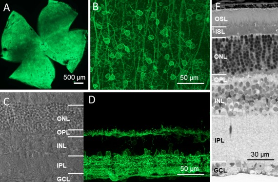
Figure 2.

Stable long-term expression of ChR2s-GFP in the retina of TKO mice. (A, B) Representative GFP fluorescence images obtained from retinal whole mounts at low (A) and high (B) magnification with the focal plane at the retinal ganglion cell layer. (C) A representative transmission image obtained from the retinal vertical section. Retinal morphology was normal, including the presence of photoreceptor cells. (D) A representative GFP fluorescence image obtained from the retinal vertical section. GFP expression was observed in cells located in the ganglion cell layer, the proximal and distal margins of the inner nuclear layer, and both the inner and outer plexiform layers. (E) Light microscope image of a semi-thin vertical retinal section. All images were obtained from mice 12 months after the injection of ChR2-C/S-GFP viral vectors. OSL, outer segment layer; ISL, inner segment layer; ONL, outer nuclear layer; OPL, outer plexiform layer; INL, inner nuclear layer; IPL, inner plexiform layer; GCL, ganglion cell layer.
