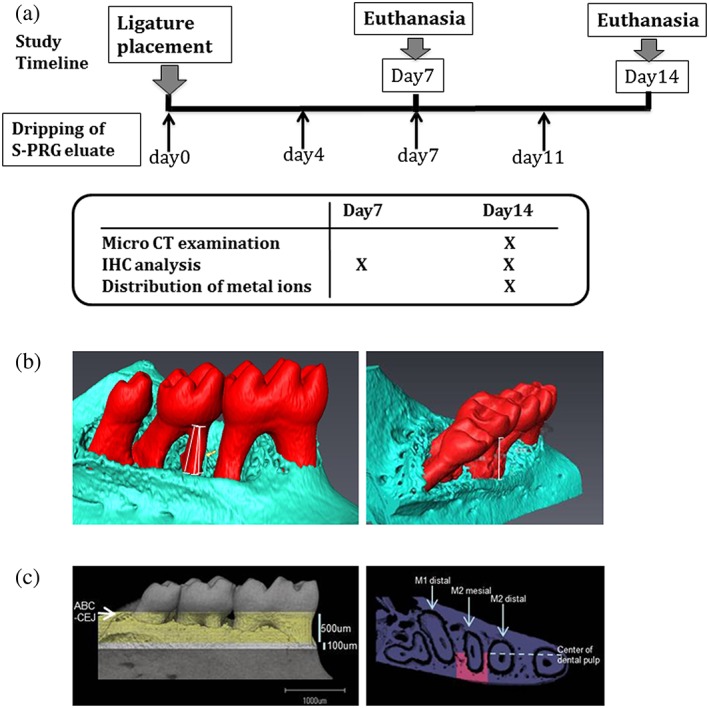
Figure 1.

Study timeline and three‐dimensional measurements in the analysis of tooth‐supporting alveolar bone. (a) The study timeline showing the time at which the surface pre‐reacted glass ionomer (S‐PRG) eluate was administered after ligature placement. Only immunohistochemical analysis was performed on day 7 and day 14. (b) A three‐dimensional view of the mandible at the site of ligature placement. Linear bone loss between the cementoenamel junction and alveolar bone crest was measured along the mesial root of M2. (c) The bone volume fraction and density around the M2 mesial root were measured in the area between 500 and 600 μm from the cementum enamel junction‐alveolar bone crest, between the distal margin of the M1 distal root and the mesial margin of the M2 distal root, and between the alveolar bone surface and the center of the dental pulp of the M2 mesial root
