
Fig. 4.
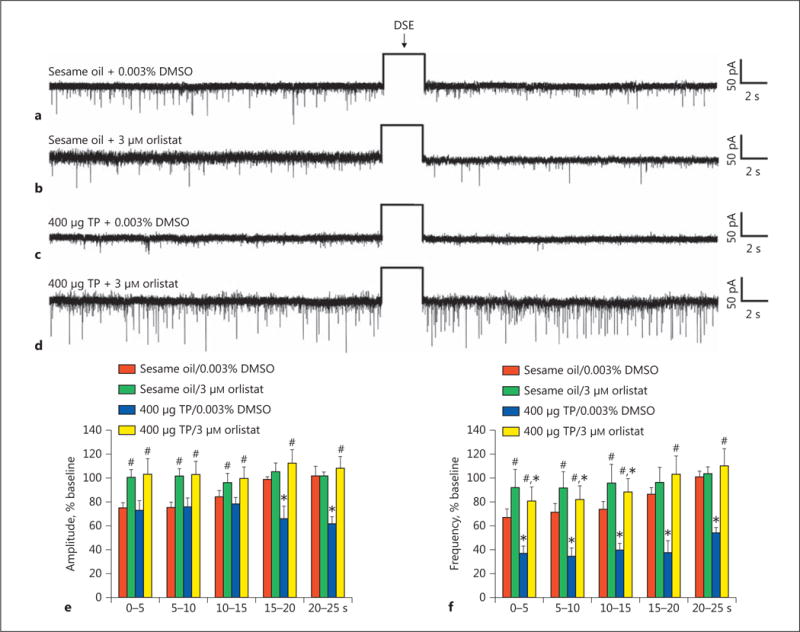
The TP-induced potentiation of DSE in identified ARC POMC neurons is abolished by orlistat. The representative membrane current traces illustrate the changes in sEPSC frequency and amplitude elicited by DSE during recordings in slices from animals treated 24 h prior with either sesame oil vehicle (0.1 mL; s.c.; a, b) or TP (400 μg; s.c.; c, d). The slices were pretreated for at least 6 min with either orlistat (b, d) or its DMSO vehicle (a, c) before electrophysiological testing. The rectangular wave under the arrow labeled “DSE” represents the truncated change in membrane current caused by the 3-s, 60-mV depolarizing voltage command. The composite bar graphs in e and f illustrate the DSE-induced changes in sEPSC amplitude and frequency, respectively, observed during the various treatment conditions. Bars and vertical lines represent means and 1 SEM, respectively. * Values of post-stimulus sEPSC frequency and amplitude observed during recordings from TP-treated animals that are significantly different (p < 0.05; rank-transformed, multifactorial ANOVA/LSD; n = 6–9) than those from vehicle-treated controls. # Values of post-stimulus sEPSC frequency and amplitude observed during recordings from orlistat-treated slices that were significantly different (p < 0.05; rank-transformed, multifactorial ANOVA/LSD; n = 6–9) than those from vehicle-treated slices.