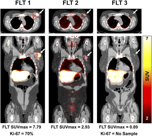
Fig. 2.

Early assessment of treatment response by FLT-PET. Fused FLT-PET/CT axial (upper) and coronal (lower) images demonstrate increased FLT uptake in an upper outer quarter breast tumor and axillary lymph node before therapy (left). After one cycle of neoadjuvant chemotherapy, there is substantial reduction in the primary breast tumor FLT uptake (middle) and resolution of FLT uptake after completion of neoadjuvant chemotherapy (right). Patient had pathologic complete response confirmed at surgery. Arrows refer to primary tumor site. This research was originally published in JNM, Ref 86. © by the Society of Nuclear Medicine and Molecular Imaging, Inc.).
