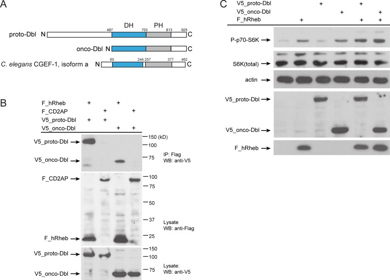
Figure 5. Human Dbl interacts with Rheb-1.
(A) Schematics of the proto-Dbl, onco-Dbl and the CGEF-1 protein domain structure. The Dbl homology (DH) domain is shaded blue, the PH pleckstrin homology (PH) domain grey. Oncogenic activation of proto-Dbl occurs through truncation of the N-terminal 497 residues. The C-terminal half of Dbl includes the DH domain and PH domain which constitutes the minimum module essential for cell transformation. (B) Dbl proteins interact with human Rheb (hRheb) in HEK293T cells. HEK293T cells expressing V5-tagged proto-Dbl or onco-Dbl and Flag-tagged hRheb-1 or CD2AP as indicated were subjected to immunoprecipitation (IP) with anti-Flag antibody. Association of V5-Dbl with Flag-hRheb was determined by western blot (WB) analysis with anti-V5 antibody (upper panel). The control protein CD2AP failed to bind Dbl. Middle part shows expression of Flag-tagged proteins, the lower panel shows the expression of V5-tagged proto- and onco-Dbl in cell lysates. kD, kiloDalton. (C) Onco-Dbl but not proto-Dbl induces S6K phosphorylation in HEK293T cells. V5-tagged proto-Dbl or onco-Dbl and Flag-tagged hRheb were co-expressed in transiently transfected HEK 293T cells. Cell lysates were analyzed by immunoblotting with the indicated antibodies. Anti-actin was used to verify equivalent input of total cellular protein.

