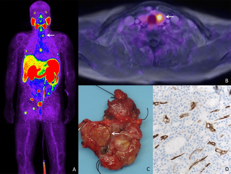
Figure 1. PSMA expression in a patient with a metastatic prostate cancer and thyroid adenoma.
(A) Bodyscan of PSMA-Pet/CT revealing uptake in several bone lesions in a patient with metastatic prostate cancer. Focal uptake is seen in the left thyroid lobe (arrow). (B) Transaxial image of fused 68 Ga-PSMA-PET/MRI showing focal uptake in a nodule in the left thyroid lobe. (C) Gross section of the thyroid lobe revealing a nodule that was histologically classified as adenoma. (D) Strong PSMA expression (200x, IHC) is shown in the adenoma-associated vasculature, apparently being responsible for the PSMA-PET/MRI uptake.

