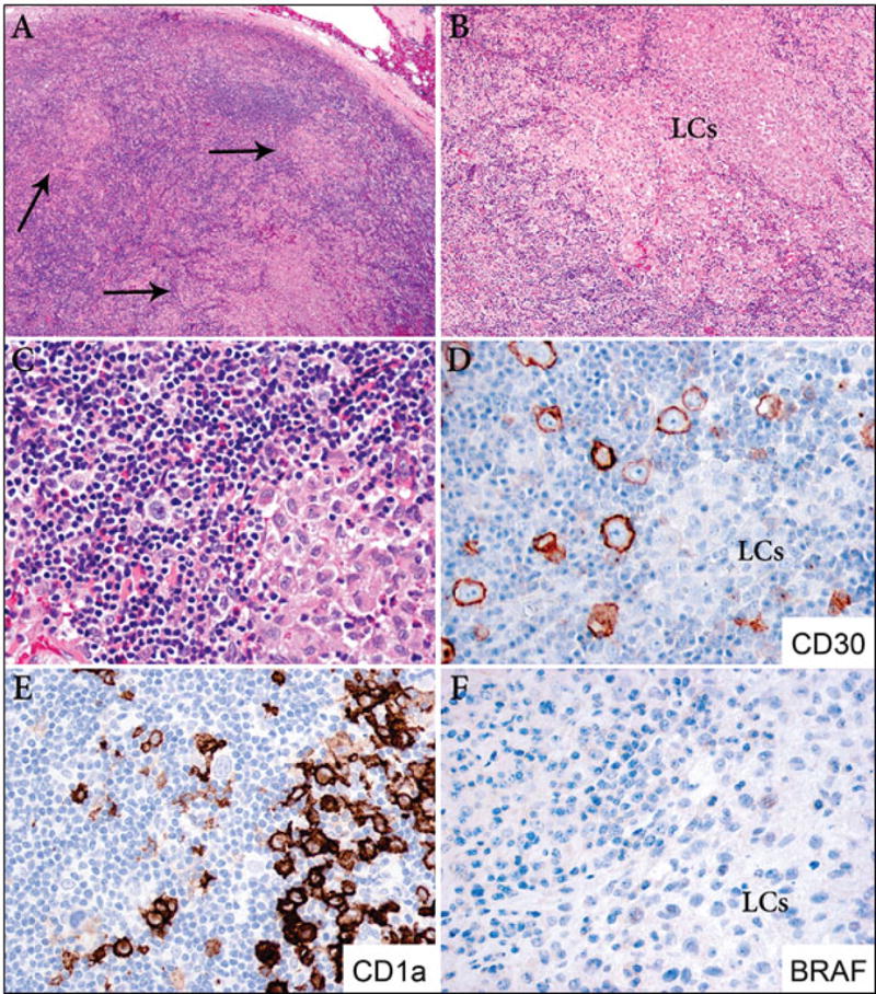
Figure 1. LCH and classical Hodgkin lymphoma, mixed cellularity (case 2).

A. Low power image of a lymph node involved by classical Hodgkin lymphoma, mixed cellularity type, and multiple clusters of LCH of variable sizes (arrows) (40X). B. Largest ill-defined nodule of LCH (200X). C. Area of transition of classical Hodgkin lymphoma with a Reed-Sternberg cell (left) and LCH (right) (400X). D. Immunostain for CD30 highlights the Reed-Sternberg cells and is negative in the Langerhans cells (LCs) (400X). E. Immunostain for CD1a is positive in the LCs and is negative in the Reed-Sternberg cells (400X). F. BRAF is negative in both components (400X).
