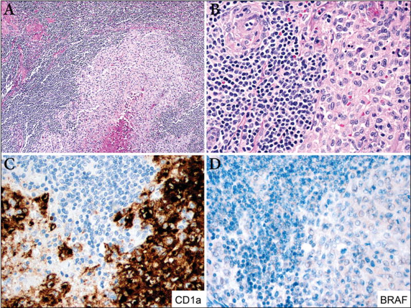
Figure 3. LCH and mantle cell lymphoma (case 7).

A. The lymph node is replaced by a monotonous proliferation of small lymphocytes with focal sclerosis. A single nodule of LCH with central necrosis is identified at the center of the lymph node (100X). B. Left, mantle cell lymphoma composed of small lymphocytes with cleaved nuclei and perivascular fibrosis. Right, LCH module with scattered apoptotic cells (400x). C. Immunostain for CD1a is positive in the Langerhans cells and is negative in mantle cell lymphoma cells (400X). D. BRAF is negative in both components (400X).
