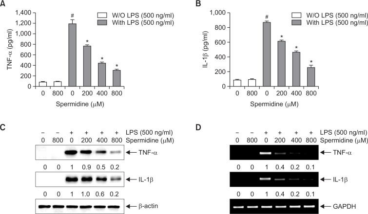
Fig. 3.
Effects of spermidine on LPS-induced secretion and expression of TNF-α and IL-1β in RAW 264.7 macrophages. The cells were pretreated with different concentrations of spermidine for 1 h, followed by stimulation with 500 ng/mL LPS for 24 h. The amounts of TNF-α (A) and IL-1β (B) in the culture supernatants were measured by ELISA kits. Data are presented as the means ± SD of three independent experiments (#p<0.05 compared to the control; *p<0.05 compared to cells cultured with 500 ng/mL LPS). (C) Cell lysates were prepared for Western blot analysis with antibodies specific for murine TNF-α and IL-1β, and an ECL detection system. (D) The total RNAs were prepared for RT-PCR analysis of the TNF-α and IL-1β mRNA expression using the indicated primers. The experiment was repeated three times, and similar results were obtained. β-actin and GAPDH were used as the internal controls for the Western blot analysis and RT-PCR, respectively. The relative ratios of expression from Western blot analysis and RT-PCR are presented at the bottom of each of the results as relative values of the β-actin and GAPDH expression, respectively.

