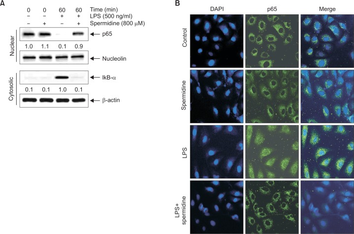
Fig. 4.
Inhibition of NF-κB nuclear translocation by spermidine in LPS-stimulated RAW 264.7 macrophages. (A) The cells were pretreated with 800 μM spermidine for 1 h before 500 ng/mL LPS treatment for 1 h. The nuclear and cytosolic proteins were prepared for Western blot analysis using anti-NF-κB p65 and anti-IκB-α antibodies, and an ECL detection system. Nucleolin and β-actin were used as the internal controls for the nuclear and cytosolic fractions, respectively. The relative ratios of expression in the results of Western blotting are presented at the bottom of each of the results as relative values of the nucleolin and β-actin expression. (B) The cells were pretreated with 800 μM spermidine for 1 h before 500 ng/mL LPS treatment. After 1 h of incubation, the localization of NF-κB p65 was visualized with fluorescence microscopy after immunofluorescence staining with anti-NF-κB p65 antibody (green). The cells were also stained with DAPI to visualize the nuclei (blue). The results are representative of those obtained from three independent experiments.

