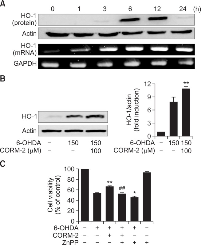
Fig. 4.
Effect of CO on expression of HO-1 in 6-OHDA-treated C6 cells (A–B) Cells were treated with 6-OHDA (150 μM) in the presence and absence with CORM-2 (100 μM) for indicated times (A) and 12 h, the peak time (B). Expression of HO-1 in either protein and mRNA levels was examined by Western blot and RT-PCR. Statistical significance was denoted by **p<0.01 compared with the 6-OHDA treatment alone. (C) A possible role of HO-1 mediating protective effect of CO against 6-OHDA-induced cytotoxicity. The cells were pre-incubated with ZnPP (0.1 μM) and then treated with 6-OHDA (150 μM) and CORM-2 (100 μM). Cell viability was examined by MTT assay. Statistical significance was *p<0.05 and **p<0.01 compared by 6-OHDA treatment alone and ##p<0.01 compared by co-treatment of 6-OHDA and CORM-2.

